Fig. 9.
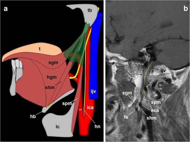
The glossopharyngeal nerve, cervical portion. a Schematic drawings that depict the glossopharyngeal nerve at the styloid pyramid and its oropharynx end. Once the Hering’s nerve (hn) has left the IX cranial nerve, the glossopharyngeal nerve enters a muscular tripod that consists of three styloid muscles and a fascia (shaded in green): the styloid pyramid. Within the pyramid, the glossopharyngeal nerve (arrowhead) usually runs in a triangular plane limited by the facial artery (*), the external carotid artery (**) and the styloglossus muscle (sgm). When the stylopharyngeus muscle merges with the constrictor muscles, the nerve enters the oropharynx and, finally, courses deep to the hyoglossus muscle (hgm). hb: hyoid bone; lc: laryngeal cartilage; shm: stylohyoid muscle; t: tongue. b Sagittal fast flair T1-weighted MRI centred at the skull base. Arrows: internal carotid artery; eca: external carotid artery; fa: facial artery; the dotted yellow line represents the expected pathway of the glossopharyngeal nerve

