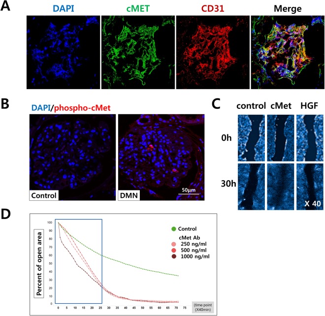
Figure 4.
Glomerular endothelial cells in vitro and wound healing analysis. (A) Immunofluorescence for DAPI (blue), cMet (green), and CD31 (red) was observed in healthy human glomeruli. Original magnification: X400. (B) The levels of phosphorylated cMet were increased in the glomeruli from patients with diabetic nephropathy compared with those from normal control subjects; DAPI (blue), phospho-cMet (red). Original magnification: X400. (C) Assessment of the endothelial cell migratory capacity was assessed using a scratch wound healing assay between 0 and 30 hours. There was a significant difference in the migratory potential of the cMet group compared to the control and HGF groups.

