Fig. 1.
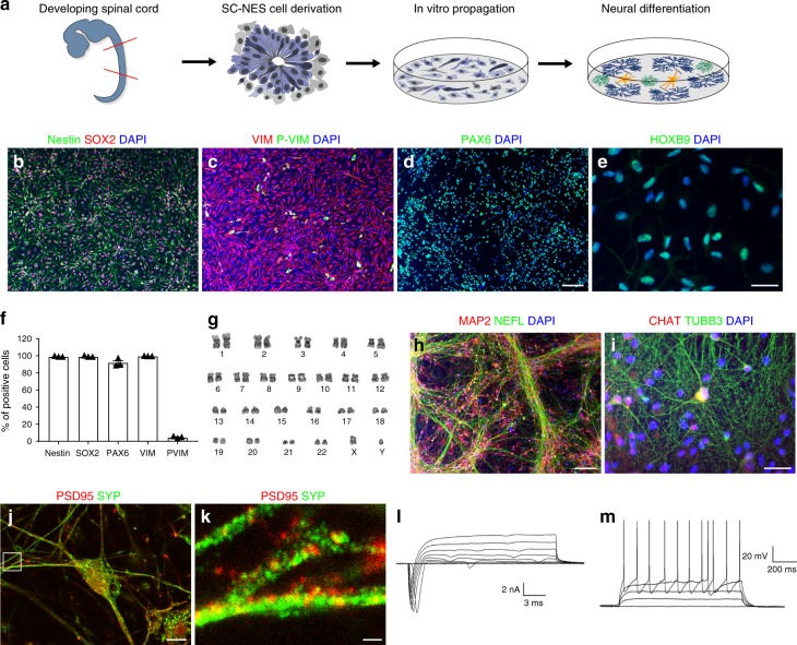
Characterization of human NES cells in proliferation and after in vitro differentiation. a Schematics of the experimental procedure. SC-NES cells were derived from the SC of a human post-mortem specimen at 6 post-conceptional weeks (PCW) and propagated in vitro. After differentiation SC-NES cells give rise to neurons, astrocytes and oligodendrocytes. b–d SC-NES cells are positive for pan-neural stem cell markers nestin, SOX2, vimentin (VIM), phospho-vimentin (P-VIM) and PAX6. e SC-NES cells retain their regional identity as proved by the positive staining for the SC-specific transcription factor HOXB9. f Quantification of nestin, SOX2, PAX6, VIM, P-VIM positive cells. g SC-NES cells retain a normal euploid karyotype after 25 passages. h, i Differentiation of SC-NES cells to MAP2-, TUBB3-, and neurofilament- (NEFL) positive neurons. SC-NES cells also generate choline acetyltransferase- (CHAT) positive neurons in agreement with their anatomical origin. j, k Mature neurons also express pre- and post-synaptic markers such as PSD95 and synaptophysin (SYP). l Total inward and outward ionic currents elicited at test potentials ranging from –70 to +40 mV from a holding voltage of –90 mV. (M) Subthreshold and suprathreshold voltage responses to a family of injected steps of current (0 pA; 20 pA; 40 pA; 60 pA; 80 pA) from a resting potential of −71 mV. Scale bars: b–d 100 µm; e 20 µm; h 100 µm; m 20 µm; j 10 µm; k 1 µm

