Fig. 3.
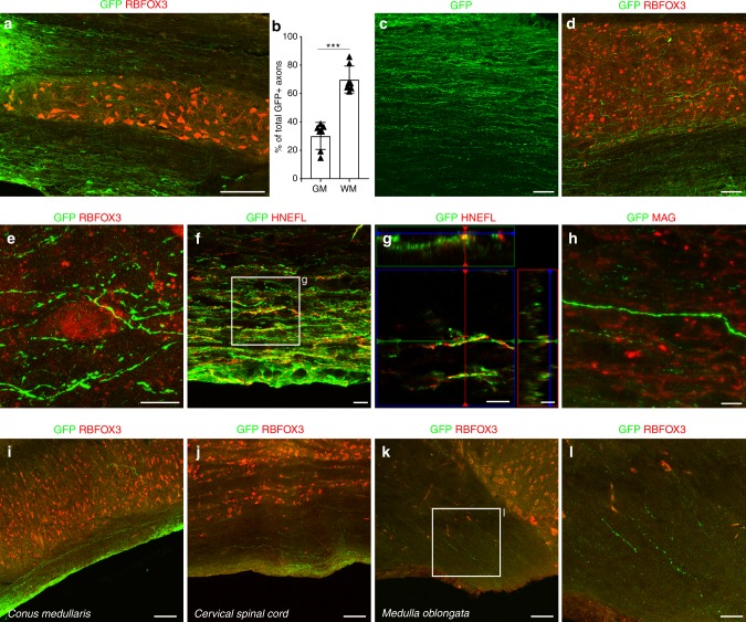
Extensive axonal outgrowth from human SC-NES cell grafts. a GFP and RBFOX3 immunolabeling on a horizontal section of SC grafted with SC-NES cells. GFP-expressing cells show a massive axonal elongation rostral and caudal to the trauma site (caudal shown). b Quantification of graft-derived axons in the gray (GM) and white matter (WM) of the recipient SC showing a preferential axonal growth in the WM. c, d GFP-positive axons grow in a parallel pattern in the WM and acquire a more ramified morphology in the GM identified by the RBFOX3 staining. e GFP-expressing axon terminals are closely associated with host RBFOX3-positive neurons. f, g GFP-labeled projections arising from the graft express human neurofilament (HNEFL), confirming their identity as axons. h GFP and myelin associated glycoprotein (MAG) staining reveals a lack of myelination of the human fibers. i–l GFP and RBFOX3 staining of the recipient SC at different levels. Human GFP-positive axonal fibers elongate throughout the entire length of the SC from the original injection site in the thoracic segment. GFP-positive axons can be found in the cervical portion and more rostrally in the medulla oblongata. Human axons also grow caudally reaching the conus medullaris. Data are expressed as mean ± s.e.m (***P < 0.001; Student’s t-test). Scale bars: a 200 µm; c, d 50 µm; e 10 µm; f–h 10 µm; i–k 100 µm; l 50 µm

