Fig. 4.
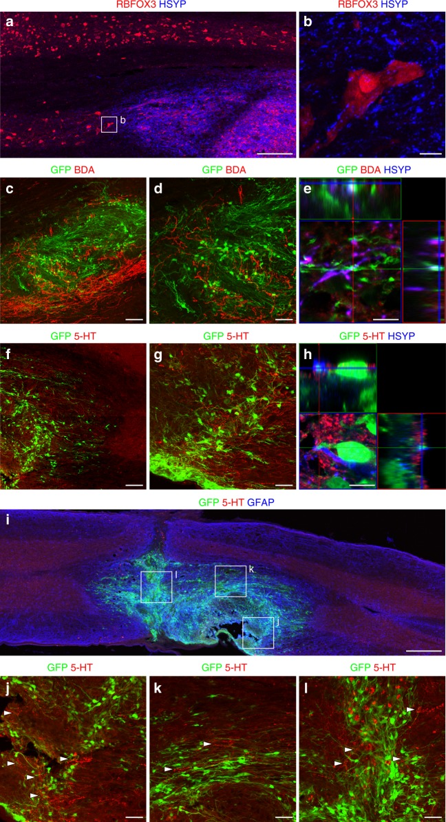
Graft-Host and Host-graft interactions. a Human synaptophysin (HSYP) staining identifies grafted SC-NES cells. RBFOX3-positive neurons indicate the gray matter of the recipient SC. b High magnification image of a host RBFOX3-positive neuron surrounded by HSYP-positive puncta, suggesting the establishment of synaptic contacts between the graft and the host. c Host corticospinal axons labeled with BDA regenerate into GFP-expressing SC-NES cell grafts. d Higher magnification of BDA positive fibers in the graft. e Adjacent signal of GFP, BDA and HSYP signal indicating the formation of novel synapses between the host CST and implanted cells. f Host serotonergic axons immunolabelled for 5-HT elongate into the GFP-positive SC-NES cell graft. g Higher magnification of 5-HT positive fibers in the graft. h Host 5-HT fibers are proximal with HSYN and GFP-positive terminals indicating the establishment of synaptic contacts between the host raphespinal fibers and grafted cells. i 5-HT-positive fibers elongate through the entire length of the cell implant. Rostral is on the right and caudal is on the left. j–l High magnification of boxed areas in (i) showing serotonergic terminals intermingled with GFP-labeled graft cells close to the rostral graft-host interface (j), in the middle of the graft (k) and in proximity of the lesion core (l). Arrowheads indicate 5-HT-positive fibers. Scale bars: a 200 µm; b 10 µm; c 100 µm; d 50 µm; e 10 µm; f 100 µm; g 50 µm; h 10 µm; i 500 µm, j–l 50 µm

