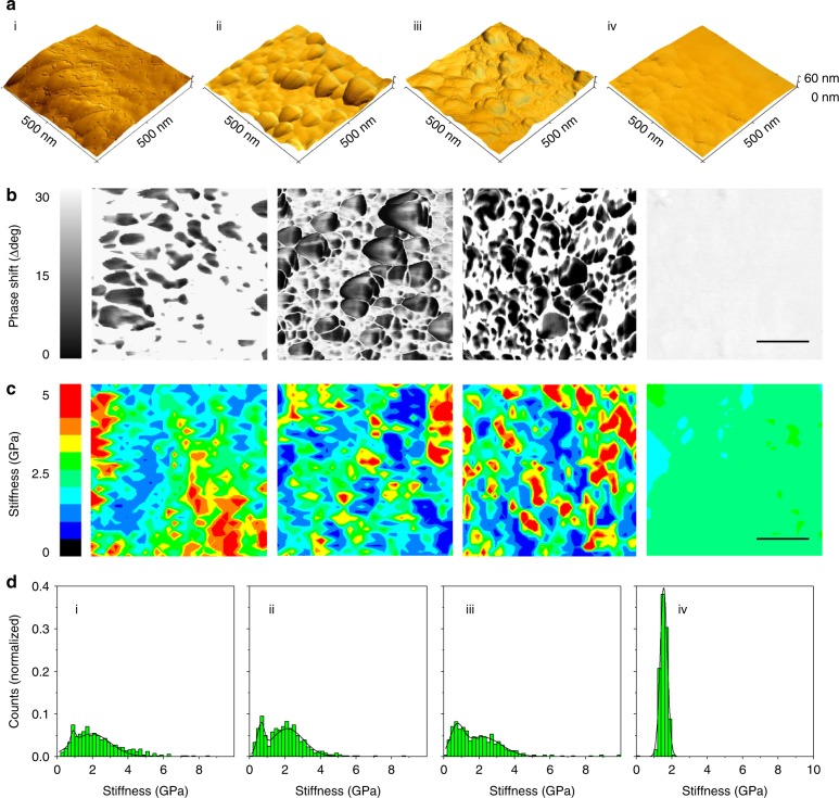
Fig. 3.
Nanomechanical investigations of the cuticle granules. All panels labeled with (i) represent M. galloprovincialis, (ii) M. californianus, (iii) S. bifurcatus, and (iv) M. capax. a High-resolution AFM topographical renderings of the abraded, exposed cuticles. b Phase shift images of AFM renderings in a (scale bar = 150 nm). c The granular structures in this channel are discernible by phase shifts to lower degrees. Corresponding color-coded force spectroscopy maps (32 × 32 pixels, scale bar = 150 nm) across these regions show a distinct link between the nanomechanical behavior and phase shifts. d Summary of binned and fitted stiffness values

