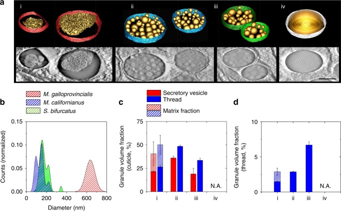
Fig. 5.
3D structural features and volume fractions. All panels labeled with (i) represent M. galloprovincialis, (ii) M. californianus, (iii) S. bifurcatus, and (iv) M. capax. a Reconstructed tomographic representations of the secretory vesicles and encapsulated structures (scale bar = 500 nm); b fitted size distributions of M. galloprovincialis (red, n = 122), M. californianus (blue, n = 176), and S. bifurcatus (green, n = 102) as deduced from TEM and electron tomographic investigations. c Granular volume fractions from the secretory vesicles (blue) compared to those of the cuticle (green, see Supplementary Figure 7 for full datasets), as well as normalized to the total thread size (d), show an increase in granular density after thread formation. Note that the intergranular matrix space in M. galloprovincialis is taken into account as well (dazzled segments, n = four tomographic sections, technical replicates, 1 µm × 1 µm × 60 Z slices, mean ± s.e.m.)

