Fig. 1.
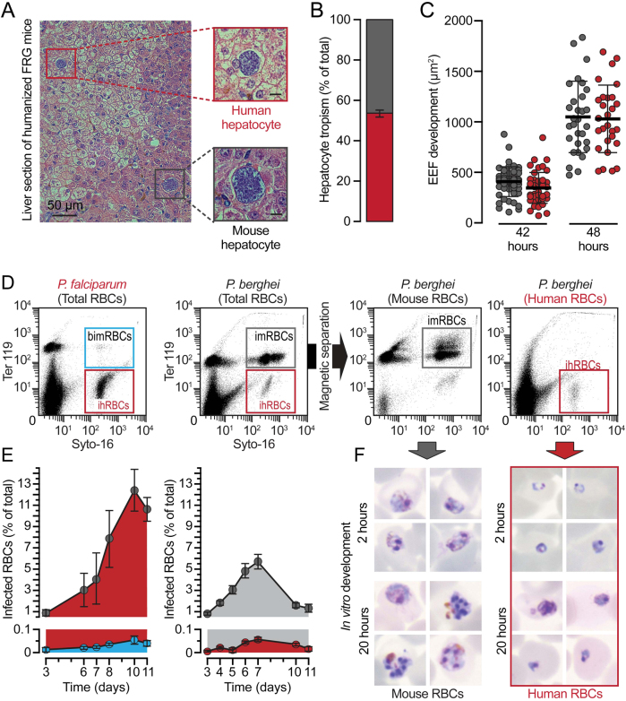
Rodent P. berghei parasites successfully develop within human hepatocytes but not within human RBCs. a Representative images of developing rodent PbWT parasites in mouse (black square) and human (red square) hepatic cells of liver-humanized FRG mice 48 h post infection (hpi) by iv injection of freshly isolated sporozoites. b Relative proportion of Pb-infected mouse (grey) and human (red) hepatocytes in humanized FRG mice, normalized to the total humanization of the chimeric liver. c PbWT development in mouse (grey) and human (red) hepatocytes 42 and 48 hpi of liver-humanized FRG mice. d Representative flow cytometry plots of peripheral blood from blood-humanized NSG mice infected by iv injection of Pf-infected (left) or Pb-infected RBCs (middle-left) before and after magnetic separation (middle-right and right); Syto-16 for nucleic acids; TER-119 for murine erythroid lineage; imRBCs/ihRBCs: infected mouse or human RBCs; bimRBCs: background signal for infected murine erythroid lineage. e Relative proportion of mouse and human RBCs infected with Pf (left) or Pb (right) parasites; bars indicate standard error. f Representative pictures of Pb parasite forms observed within magnetically separated imRBCs and ihRBCs from the total blood of infected blood-humanized NSG mice after 2 and 20 h of in vitro culture

