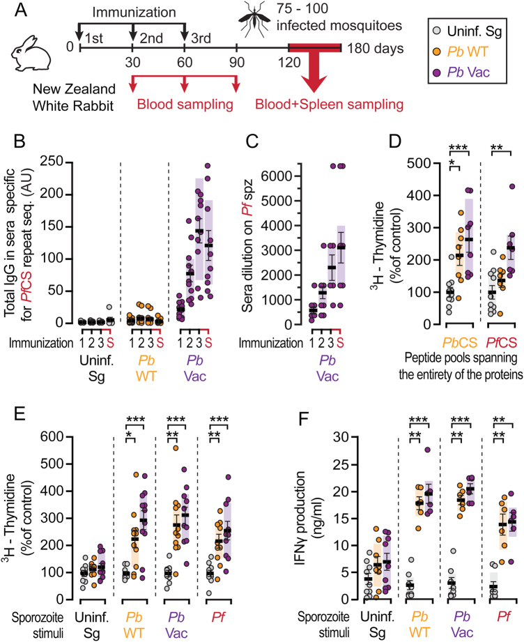
Fig. 4.
Immune responses in NZW rabbits after PbVac sporozoite immunization. a Diagram of the immunization protocol. Immunizations were performed by exposure to the bites of 75-100 mosquitoes. b Total IgG titers against PfCS repeat sequence in serum after 1, 2, and 3 mock immunizations (grey), or immunization with PbWT (orange) and PbVac (purple), or at the time of animal sacrifice (S) 60–90 days after last immunization. c Serum binding capacity to Pf sporozoites after PbVac immunization. d Spleen cell proliferation upon stimulation with peptide pools spanning the entire PbCS or PfCS proteins in immunized rabbits, as indicated by assessment of 3H-thymidine incorporation. e Spleen cell proliferation upon stimulation with PbWT, PbVac or Pf sporozoites, as indicated by assessment of 3H-thymidine incorporation. Stimulation with an extract of uninfected mosquito salivary gland material was used as control. f IFNy production in rabbit spleen cell supernatant after stimulation with PbWT, PbVac or Pf sporozoites. Measurements were taken from distinct samples. The boxes correspond to the 25th and 75th percentiles; the line and bars indicate mean of infection and standard error of the mean, respectively; *p < 0.05; **p < 0.01; ***p < 0.001, as determined by Kruskal–Wallis test, corrected with Dunn’s multiple comparisons test

