Fig. 4.

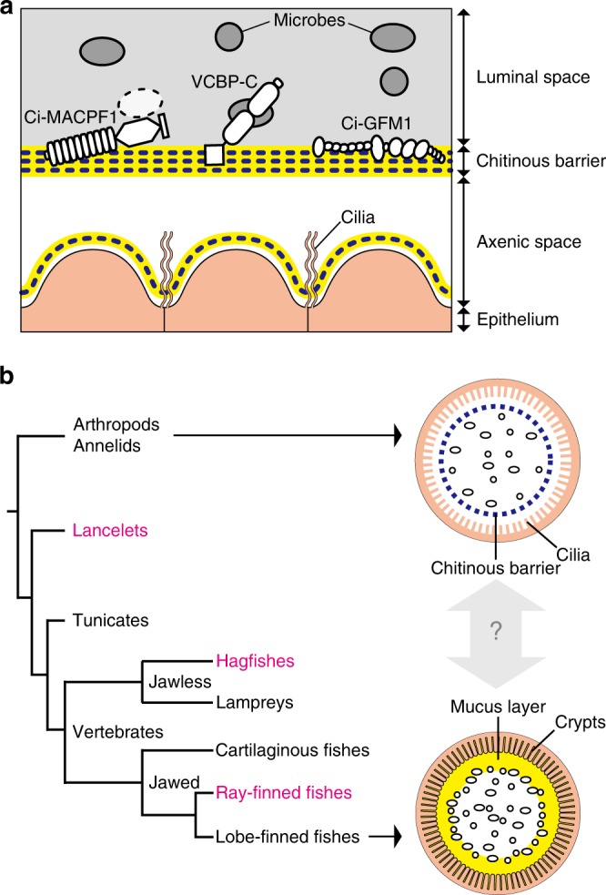
A chitin-based barrier immunity model. a A model. The intestinal mucosal surface of the tunicate, C. intestinalis Type A, is axenic due to the barrier function of multi-layered chitinous membranes that confines microbes to the luminal space. The barrier function results from sieving by a chitinous framework (blue dotted lines) and immune functions of matrix substances (yellow lines), e.g., cytolytic Ci-MACPF1 (left), bacteria-seizing VCBP-C (center), and possible multimeric protein Ci-GFM1 (right). Delamination of a new membrane from the epithelium renews axenic conditions. b Evolutionary implications. The tree diagram depicts phylogeny of chordates (lancelets, tunicates and vertebrates) and invertebrate outgroups (arthropods and annelids). Two arrows extending from outgroups and lobe-finned fishes point to schematic drawings of intestinal barrier immunity representative of each group. Note that these simple drawings highlight physical, but not cellular nor chemical components of barrier immunity. Invertebrate outgroups share a chitinous barrier membrane, known as the peritrophic matrix (PM, a blue dotted circle)21,32, which encloses food matter and luminal microbes (ovals). The mammalian subgroup of lobe-finned fishes possesses a GFM-based mucus layer (a yellow circle) that covers the mucosal surface and hampers microbial access to the epithelium, while harboring dense microbes (ovals)24,31. The second diagram shows the distal colon of mice. The mammalian mucus system has multiple physiological roles, and the condition of mucus varies along the longitudinal axis of the intestine. Although invertebrate PMs and mammalian mucus layers are considered analogous, i.e., with no common descent, the chitin-based barrier immunity newly found in tunicates (a) provides molecular evidence for a possible link between these two barrier immune systems. To test this idea, animal groups that occupy intervening phylogenetic positions (typed in magenta) are critical
