Fig. 6.
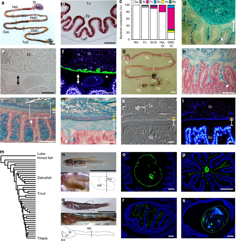
Microbial colonization is a major distinction between the mucus layers of ray-finned fish and mice. a Gastrointestinal tract of O. mossambicus. St. stomach, HeL hepatic loop, PMC proximal major coil, GaL gastric loop, DMC distal major coil, TeS terminal segment. b In situ hybridized cross-section of DMC, showing expression of a chitin synthase gene Om-CHS1 in the epithelium (Ep). LaP lamina propria, Lu lumen. c Intestines harbor indigenous bacterial community. This panel summarizes 16S rRNA gene analysis of aquarium water (Wa), food (Fo), stomach digesta (St-Di), HeL digesta (HeL-Di), and DMC digesta (DMC-Di). Ga gammaproteobacteria, Al alphaproteobacteria, Fu fusobacteria, Ba bacteroidetes, Ve verrucomicrobia, Be betaproteobacteria. For details, see Supplementary Table 2. d Alcian blue-stained cross-section of DMC. Goblet cell-derived mucus fills the space between digesta (Di) and the epithelium (double-headed arrow). Digesta contains abundant mucus from pharyngeal regions. An arrowhead denotes a goblet cell. e, f Cross-sections of DMC (e Phase-contrast; f CBD-DAPI double-staining). A chitinous membrane (green) separates digesta microbes (blue) from the mucus layer covering the DMC epithelium (double-headed arrows). An arrowhead denotes a DAPI signal at the surface of the mucus layer. g Gastrointestinal tract of a mouse. Du duodenum, Je jejunum, Il ileum, Ce cecum, Co colon, Re rectum. h–j Alcian blue-stained gut sections counterstained with nuclear fast red (h, ileum; i, colon; j, the boxed area in i). Colon mucus covers the epithelium and consists of an inner layer devoid of microbes (white) and an outer layer densely colonized with microbes (yellow). Arrowheads show mucus granules in goblet cells. k–l Cross-sections of colon (k, DIC; l, DAPI). m Phylogeny of ray-finned fish65, showing lobe-finned fishes (outgroup), zebrafish, rainbow trout and tilapia. n Zebrafish secondarily lost the stomach. The anterior intestine enlarges as the intestinal bulb (InB). PoI posterior intestine. o, p CBD-DAPI double-stained sections at the dotted line in n (o, InB; p, PoI). q Rainbow trout fry. The stomach (St) bends anteriorly, followed by the intestine. AnI anterior intestine, MiI middle intestine. r, s CBD-DAPI double-stained sections at dotted lines in q (r, AnI; s, MiI). For clarity, liver, gall bladder and spleen were removed in n and q. Scale bars (a, g, n, q) 1 cm, (b, d–f, h, i, o, p, r, s) 100 μm, and j–l 20 μm

