Figure 4.
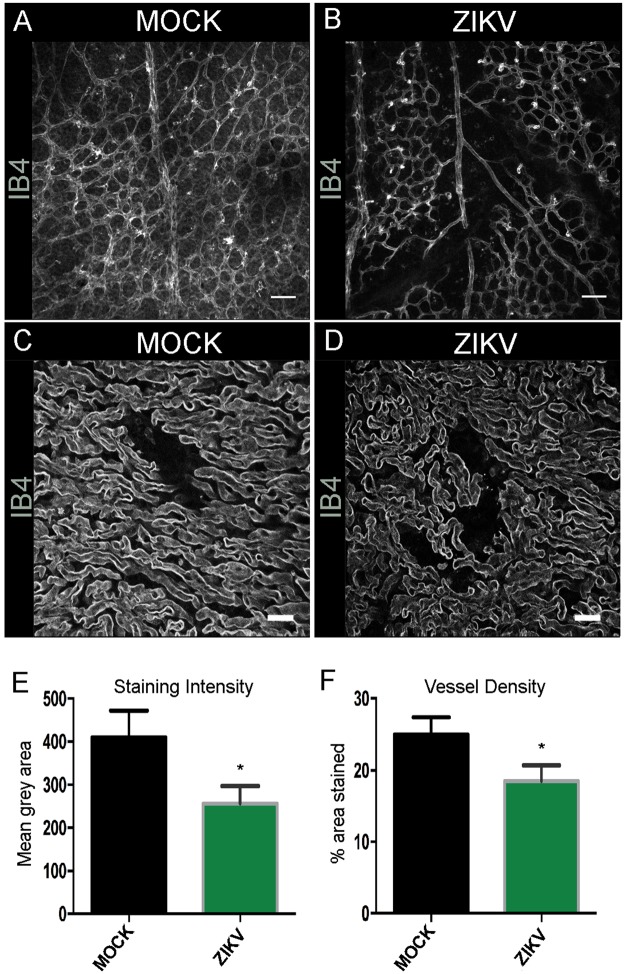
ZIKV infected placental vessel morphology at E15.5 Confocal images of IB4 stained wholemount retinas at P7 MOCK (A) and ZIKV infected animals (B). Scale bars = 100 μm. n = 7. Confocal images of IB4 stained blood vessels in 50 μm placenta section at E15.5 MOCK (C) and ZIKV infected animals (D). Scale bars = 20 μm. We analysed three sections from at least three different placentas for each condition. Blood vessel staining intensity (E) and vessel density (F) were measured with ImageJ and found reduced in ZIKV infected placenta compared to MOCK at E15.5. Student’s t-test *p < 0.05.

