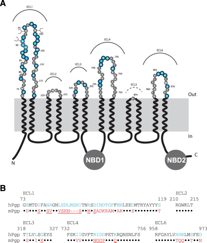
Figure 1.
Topology and sequence comparison of the extracellular loops of human and mouse P-gp. (A) Schematic representation of the primary structure of human P-gp. ECLs 1 to 6 are labeled. Residues that are different in human and mouse P-gps are highlighted in light blue. (B) Sequence alignment of putative human and mouse P-gp extracellular loop residues. The ECL5 was omitted due to its shortness and sequence conservation. Residues M89, S90 and N91 are not present in mouse P-gp. The residues that are different in human and mouse P-gp are shown in blue and red, respectively. Conserved residues in both human mouse transporters are presented as black dots. The mutated residues in the ECL regions of mouse P-gp are underlined.

