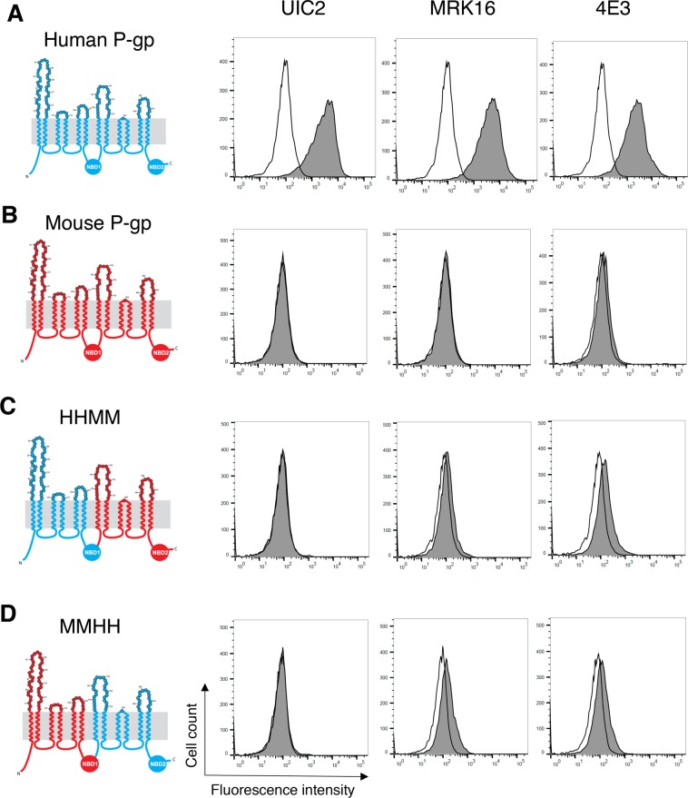
Figure 2.
Extracellular loops from both halves of human P-gp are required for binding of the three antibodies. Schematic of the 2-D topology of P-gp (left) and representative histograms of flow cytometry analysis of antibody binding to WT human, mouse and different human-mouse P-gp chimeras are shown (right). WT human P-gp (A), WT mouse P-gp (B), HHMM (C), and MMHH (D) chimeras. The amino acids in the extracellular loops are color coded. Human-specific and mouse-specific amino acids are shown in blue and red, respectively. HeLa cells transduced with BacMam baculovirus carrying WT human, WT mouse, HHMM or MMHH P-gp chimeras were harvested 24 hours post-transduction and incubated at 37 °C with human P-gp specific antibodies (at indicated concentration per 100,000 cells) UIC2 (2 µg), MRK-16 (1 µg), and 4E3 (1.5 µg) (filled gray traces) or IgG2a control isotype (2 µg) (unfilled traces). Following incubation with primary antibodies, the cells were washed and incubated with FITC-conjugated secondary antibody at 37 °C for 30 min and the antibody binding was measured by flow cytometry (compare filled grey and unfilled traces in histograms). Similar results were obtained in three or more independent experiments.

