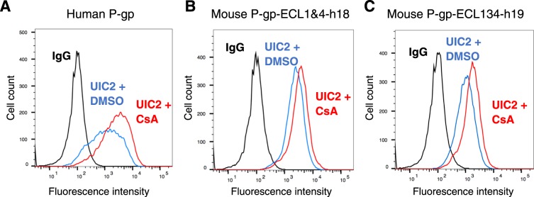
Figure 6.
Substrate-dependent conformational changes in UIC2 binding to mouse-human P-gp chimeras are the same as in human P-gp. (A) WT human P-gp, (B) mouse P-gp-ECL1&4-h18 and (C) mouse P-gp-ECL13&4-h19 chimera-expressing HeLa cells were incubated for 5 minutes at 37 °C with DMSO (solvent control) or 20 µM cyclosporine A before adding UIC2 antibody (at saturated concentration, 3 µg/100,000 cells). After 30 min incubation, the cells were washed and incubated with FITC-labeled anti-mouse secondary antibody for another 30 min before acquiring data with flow cytometry. Histograms from a typical experiment are shown. The experimental details are given in each histogram. The blue traces show the UIC2 binding in the presence of DMSO and red traces depict antibody binding to cells pretreated with cyclosporine A (UIC2 shift assay). Black traces correspond to binding to the IgG control. Similar results were obtained with two additional independent experiments.

