Figure 4.
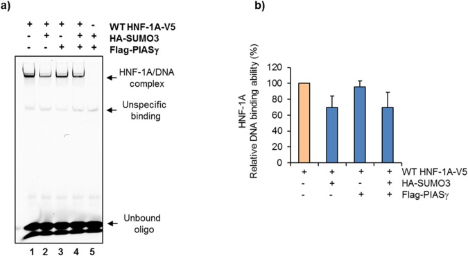
PIASγ does not influence the DNA binding ability of HNF-1A. (a) The DNA-binding ability of HNF-1A was assessed in nuclear fractions isolated from HeLa cells transiently transfected with WT HNF-1A or empty vector, and co-transfected with SUMO-3 and PIASγ. HNF-1A-DNA binding was analyzed by electrophoretic mobility shift assay (EMSA) after HNF-1A incubation with a Cy5 labeled DNA oligo (corresponding to HNF-1 binding site in the rat albumin). Bound complexes were separated on a 6% DNA retardation gel and the fluorescence signal was detected at 670 nm. Full-length gel is presented in Supplementary Fig. S6. (b) Quantification of binding by densiometric analysis is presented relative to WT alone (set to 100%). Each bar represents a mean of three independent experiments ±SD (n = 3).

