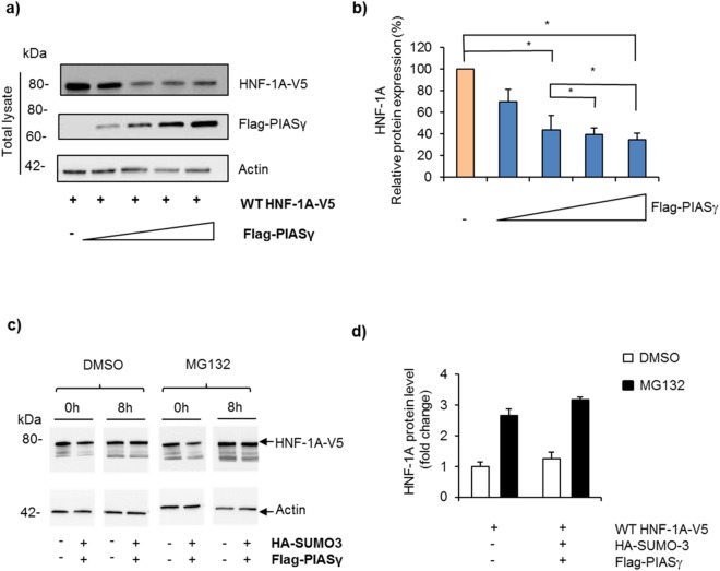
Figure 7.
PIASγ reduces the cytosolic HNF-1A protein level in a dose- dependent manner. (a) Effect of PIASγ on HNF-1A protein in cleared lysates from transiently transfected HEK293 cells, with V5-tagged HNF-1A together with increasing amounts of Flag-tagged PIASγ (0.5–2 µg), and analyzed by SDS-PAGE and immunoblotting using indicated antibodies. Full-length blots are presented in Supplementary Fig. S9. (b) The level of HNF-1A was normalized to actin and presented relative to the level of HNF-1A alone. The results are shown as mean of four independent experiments ±SD (n = 4). *Indicates p < 0.05. (c) HEK293 cells were transiently transfected with V5-tagged HNF-1A in the presence or absence of HA-SUMO-3 and Flag-tagged PIASγ. Post-transfection, cells were treated with 10 µg MG132 or DMSO for 8 hours, and protein from cleared lysates was analyzed by SDS-PAGE and immunoblotting using indicated antibodies. Full-length blots are presented in Supplementary Fig. S10. (d) Quantification of proteins shown in c) by densiometric analysis. The level of HNF-1A was normalized to the loading control (actin). Each column represents the mean fold difference in the MG132-treated samples versus DMSO-treated control on two different experimental days ± SD (n = 2).

