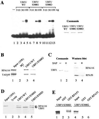
Figure 5.
Phosphorylation at Ser-388 is necessary for association of UBF1 with Pol I. (A) Mutation of Ser-388 does not interfere with DNA binding of UBF. A 32P-labeled cruciform DNA probe was incubated with 3, 10, 30, and 100 ng of FLAG-tagged UBF1/WT (lanes 2–5), UBF1/S388G (lanes 6–9), and UBF1/S388D (lanes 10–13) immunopurified from baculovirus-infected Sf9 cells. UBF-DNA complexes were analyzed on a 6.5% PAA-gel. A Coomassie blue stain of 500 ng of UBF1/WT, UBF1/S388G, and UBF1/S388D is shown at Right. (B) Pull-down experiment. Extracts from FM3A cells were incubated for 1 h at 4°C with either bead-bound FLAG-tagged UBF1/WT, UBF1/S388G, or control beads saturated with the FLAG-epitope peptide as indicated. Bead-bound proteins were analyzed on Western blots by using antibodies against TAFI95 and RPA116. Lane 1 shows 10% of input TAFI95 and RPA116. (C) Phosphatase treatment impairs the interaction between Pol I and UBF1. Bead-bound FLAG-UBF1/WT was preincubated in the absence or presence of shrimp alkaline phosphatase (SAP) for 30 min at 30°C. Bead-bound UBF1 was analyzed by Coomassie staining (lanes 1 and 2) and assayed for interaction with Pol I by using antibodies against RPA116 and RPA53 (lanes 3 and 4). (D) Coimmunoprecipitation experiment. Fifty microliters of Pol I (H-400 fraction) were incubated with 35S-labeled FLAG-tagged UBF1/WT (lane 3), UBF1/S388G (lane 4), or UBF1/S388D (Lane 5), followed by immunoprecipitation with M2− antibodies. An unprogrammed reticulocyte lysate was used as a control (lane 2). UBF was visualized by autoradiography, Pol I was monitored on Western blots by using α–RPA116 antibodies. Lane 1 shows 10% of RPA116 present in the fraction. (E) Interaction between UBF and RPA53. 35S-labeled UBF1/S388D (lanes 1–3) or UBF1/S388G (lanes 4–6) were incubated with immobilized GST (lanes 2 and 5) or GST-RPA53 (lanes 3 and 6). Bead-bound proteins were separated on 8% SDS/PAA gels and visualized by autoradiography. Lanes 1 and 4 show 10% of the UBF used.

