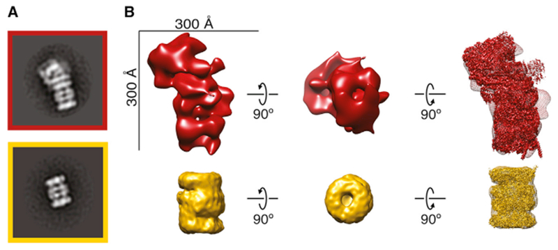
Figure 5. Ab Initio Structures from a Cellular Fraction Unambiguously Reveal the Proteasome.

(A) Reference-free 2D class averages of the proteasome from Figure 3B.
(B) Top: structure of single-capped proteasome generated using RELION from manually picked particles. Bottom: ab initio structure of the 20S core proteasome generated using cryoSPARC is shown. High-resolution structures EMD-4002 (Schweitzer et al., 2016) and EMD-2981 (da Fonseca and Morris, 2015) are fit into the structures, respectively.
See also Figures S4 and S5.
