
Figure 3.
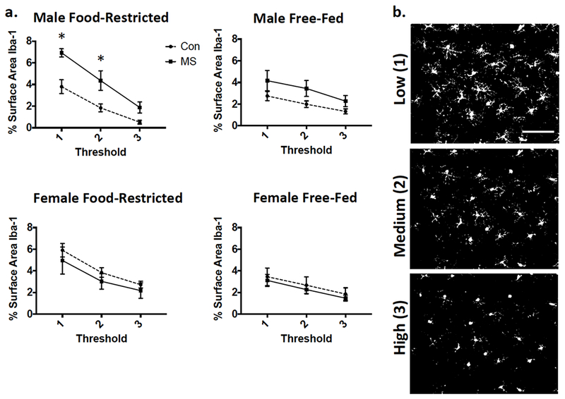
Iba-1 immunofluorescence as expressed by % surface area at each of three thresholded analyses. (a) Graphical representation of means ± SEM (n=9-12). Con: control-reared; MS: maternally separated. (b) Examples of analyzed images after threhsolding at each level. Images taken from a control-reared, food-restricted male. Scale bar: 100μm