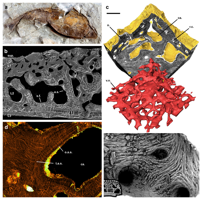
Figure 2. Morphology and histology of the heterostracan dermal skeleton.
Gross external morphology of the dermal skeleton of Errivaspis waynensis (NHMUK P19789) from the Lochkovian of Herefordshire, UK (a); etched SEM section of Loricopteraspis dairydinglensis (NHMUK P75400) showing the 4-layered construction of the dermal skeleton. Aspidin spaces and Sharpey’s Fibre spaces are preserved as high relief pyrite diagenetic infill. Sharpey’s Fibre spaces pervading L3 can be distinguished from aspidin spaces in L2 by both their size and configuration (b); sectioned srXTM virtual model of the dermal skeleton ofTesseraspis tesselata (NHMUK P73617). The vasculature network is shown to comprise a series cancellae interlinked by reticular canals (c); SrXTM horizontal virtual thin section through aspidin trabeculae ofTesseraspis tesselata (NHMUK P73618). Aspidin spaces are preserved as diagenetic pyrite infill with high X-ray attenuation. Spaces are organised orthogonal to the trabecular lamellae, or else tangled at trabecular intersections (d); SEM detail of a cancellar chamber of Loricopteraspis dairydinglensis (NHMUK P73622), showing a centripetal fabric of coarse spicules, interpreted as mineralised (crystal) fibre bundles (e); SrXTM horizontal virtual thin section through aspidin trabeculae of Tesseraspis tesselata (NHMUK P73618). Aspidin spaces are preserved as diagenetic pyrite infill with high X-ray attenuation. Spaces are organised orthogonal to the trabecular lamellae, or else tangled at trabecular intersections (e). r.c., reticular canal; o., odontode; v.n., vascular network; ca, cancellae; a.s., aspidin space; o.a.s., orthogonal aspidin space; t.a.s., tangled aspidin space; s.f., Sharpey’s Fibre space; sup, superficial layer; L1, layer 1; L2, layer 2; L3, layer 3. Relative scale bar equals 21mm in (a), 183 μm in (b), 165 μm in (c), 83 μm in (d). and 49 μm in (e).

