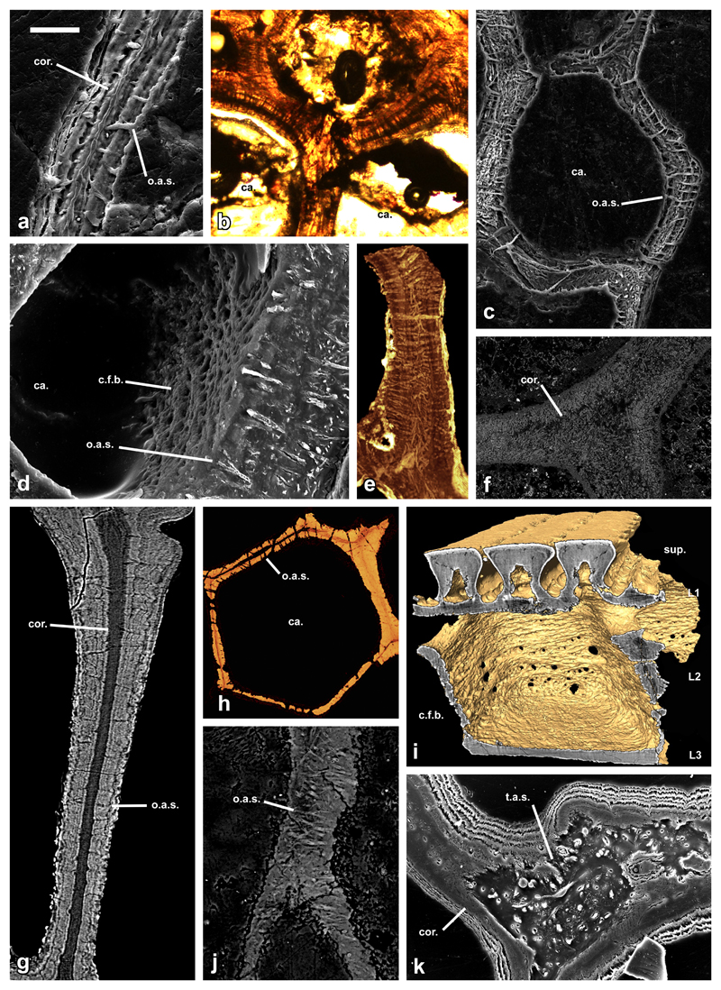
Figure 3. Histology of aspidin in phylogenetically disparate heterostracan taxa.
SEM etched section of a trabecular wall of Lepidaspis serrata NRM-PAL C.5940 showing bipartite construction (a); LM thin section of a junction of trabecular walls of Phialaspis symondsi NHMUK P73619 (b); etched SEM section through a polygonal cancellar chamber of Corvaspis kingi NHMUK P73616 (c); BSE SEM vertical section through a polygonal cancellar chamber of Corvaspis kingi NHMUK P73613 showing both the aspidin spaces and rystal fibre bundles (d); horizontal SrXTM virtual thin section through trabecular walls of Tesseraspis tesselata NHMUK P73617 (e); SEM BSE section of a trabecular junction of Amphiaspis sp. GIT 313-32 (f); SrXTM tomographic slice through a vertical trabecular of Anglaspis macculloughi NHMUK P73620 (g); SrXTM horizontal virtual thin section through a polygonal cancellar chamber of Pteraspis sp. NRM-PAL C.5945 (h); isosurface model of the same specimen showing the circular fabric of fibres enveloping the cancellae (i); vertical trabecular wall of Poraspis sp. NHMUK P17957 (j); junction of trabecular walls of Loricopteraspis dairydinglensis NHMUK P73623 (k). sup, superficial layer; L1, layer 1; L2, layer 2; L3, layer 3; ca, cancellae; o.a.s., orthogonal aspidin space; t.a.s., tangled aspidin space; c.f.b., crystal fibre bundles; cor., homogenous core of bipartite aspidin trabeculae. Relative scale bar equals 30 μm in (a), 48 μm in (b), 79 μm in (c), 30 μm in (d), 88 μm in (e), 180 μm in (f), 41 μm in (g), 100 μm in (h), 91 μm in (i), 40 μm in (j) and 52 μm in (k).

