
Fig. 6. Effect of PBM treatment on caspase-9 and caspase-3 activity and apoptotic cell death induced by HI.
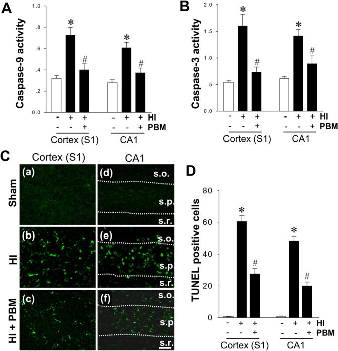
A, B The change of caspase-9 and caspase-3 activity in the proteins from cortex (S1) and hippocampal CA1 was examined using specific AMC-based fluorometric substrates at P18 from sham and HI rats. The fluorescence of free AMC was measured and compared between groups. C Representative confocal microscopy images depict fluorescent TUNEL staining (green) in cortex (S1) and hippocampus CA1 region (s.o., stratum oriens; s.p., stratum pyramidal and s.r., stratum radiatum). Scale bar: 20 µm. D The numbers of TUNEL positive cells from the indicated groups were counted and statistically determined. Results are means ± SE from 4–5 animals in (A) and (B), and 8–12 animals in (C) and (D) in each group. *P < 0.05 versus sham, #P < 0.05 versus HI control group.