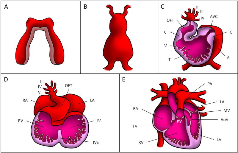
Figure 1. Cardiac formation stages.
(A) Cardiogenic cords, (B) linear tubular heart, (C) looped tubular heart, (D) cardiac septation, and (E) fully formed four-chambered heart. OFT: outflow tract, AVC: atrioventricular canal, C: endocardial cushion, V: primitive ventricle, T: trabeculae, A: primitive atrium, RA: right atrium, LA: left atrium, RV: right ventricle, LV: left ventricle, IVS: interventricular septum, PV: pulmonary valve, PA: pulmonary artery, MV: mitral valve, AoV: aortic valve, TV: tricuspid valve. The roman numbers in (C) and (D) correspond to the numbers assigned to the pharyngeal arch arteries (6 in total over developmental stages).

