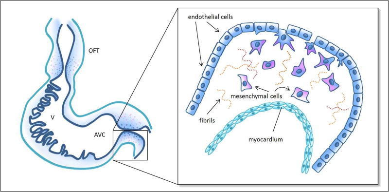
Figure 2. Schematics of endothelial-to-mesenchymal transition (EMT).
Left: schematic representation of the looped tubular heart depicting cushions in the outflow tract (OFT) and atrioventricular canal (AVC), as well as trabecular structures in the primitive ventricle (V). Inset: details of EMT in the AVC cushions, wherein a subset of activated endothelial cells (navy) delaminate from the endocardium, elongate, develop filopodia, migrate into the cardiac jelly, which is composed of extracellular matrix including some fibril proteins, and acquire a mesenchymal phenotype (pink). See Table 1 for relevant developmental periods.

