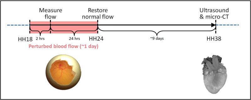
Figure 5. Schematics of experimental design.
Blood flow was perturbed at HH18 through VVL or OTB, and resulting blood flow dynamics were measured 2 hours after intervention. Embryos were then reincubated to HH24 (~24 hours after intervention) and either collected for further analysis or had their hemodynamics restored (for OTB embryos). Embryos that were not collected were then re-incubated and analyzed for structural cardiac malformations at HH38, when the heart was fully formed.

