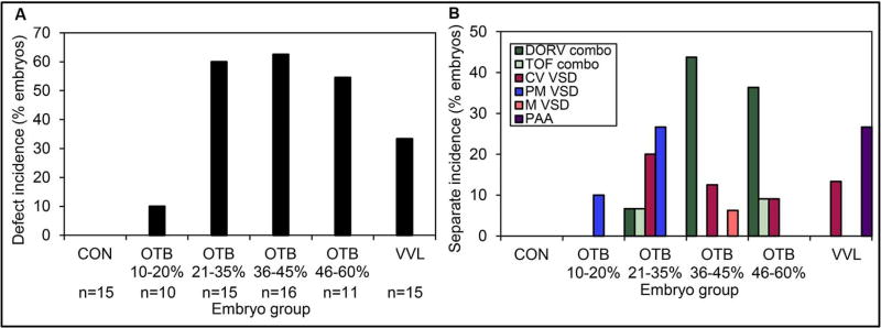
Figure 8. Cardiac defects depend on the level of hemodynamic perturbation.
(A) Overall defect incidence among surviving embryos. Embryos were grouped by band tightness, VVL intervention, and controls were also included. (B) Separate defect type incidence among surviving embryos, by embryo group. CON: normal control, OTB: outflow tract banded embryos, VVL: vitelline vein ligated embryos, VSD: ventricular septal defect, CV VSD: conoventricular VSD, PM VSD: perimembranous VSD, M VSD: muscular VSD, DORV: double outlet right ventricle, TOF: Tetralogy of Fallot. Reproduced from (Midgett et al., 2017b).

