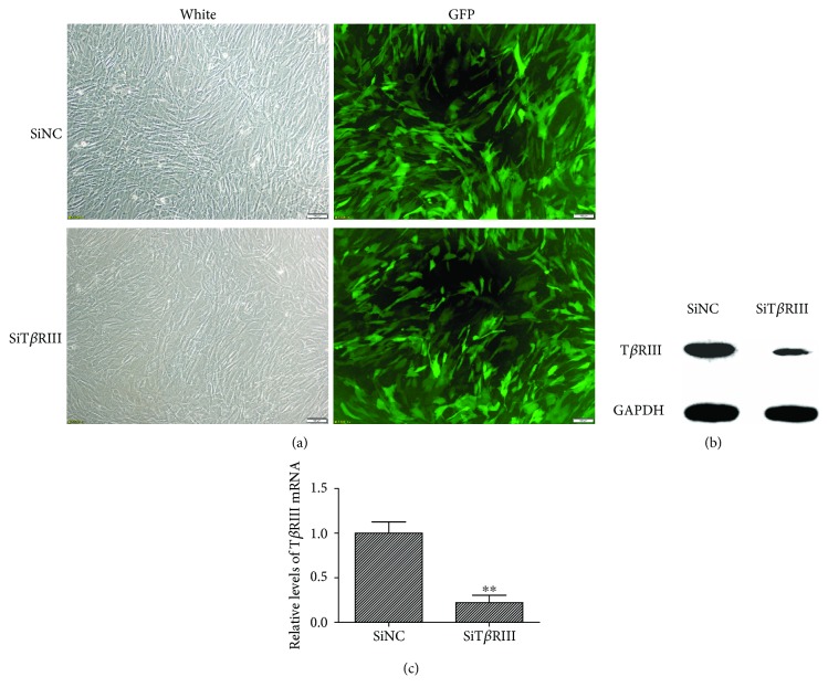
Figure 2.
Identification and efficiency of lentiviral infection. (a) GFP fluorescence imaging confirmed that the majority of hMSCs were GFP positive 72 h after they were infected by TβRIII siRNA (SiTβRIII) or SiNC virus. Scale bar = 100 μm. (b) Western blot showed that TβRIII siRNA clearly inhibited the expression of the TβRIII protein. (c) qPCR confirmed that the expression profiles of TβRIII mRNA decreased significantly in the SiTβRIII groups as compared with those in the SiNC group. Error bars represent the means ± SD, n = 3. ∗∗P < 0.01 versus SiNC group.

