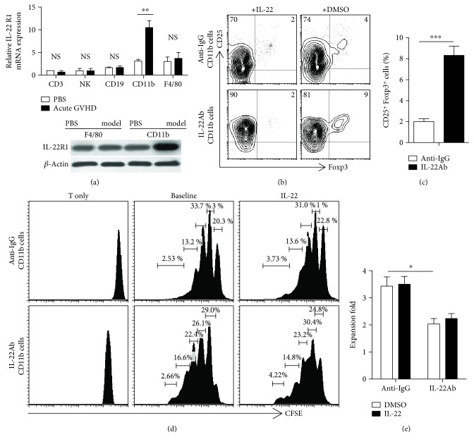
Figure 5.
IL-22Ab treatment increased Foxp3 expression and suppressed T cell proliferation via CD11b+ cells. CD11b+ cells were obtained from the spleen of anti-IgG- or IL-22Ab-treated aGVHD mice by positive selection using anti-PE-CD11b and anti-PE beads (Miltenyi Biotec). CD4+CD25− T cells were obtained from the spleen of normal B6 mice. The two cell subsets were cocultured at a ratio of 1 : 5 for 3 days. In some experiments, IL-22 was replenished to verify its function in T cell differentiation and expansion. (a) Relative expression of IL-22R1 mRNA in CD3+ T cells, natural killer (NK) cells, CD19+ cells, CD11b+ cells, and F4/80+ cells in normal D2B6F1 mice and aGVHD mice (day 7). The CD3 cell vehicle of the PBS group was arbitrarily assigned a value of 1, with other values presented relative to this value. The expression of IL-22R1 on CD11b+ and F4/80+ cells derived from aGVHD mice on day 7 was determined by Western blotting. (b) Representative contour plots of CD25+Foxp3+ cells with the addition of TGF-β and IL-2; cells were gated on CD4+ T cells 3 days after culture. DMSO: dimethyl sulfoxide. (c) Relative number of CD25+Foxp3+ T cells among CD4+ cells. (d) Representative histogram plots of T cell expansion after coculture with aGVHD model-derived CD11b+ cells in the presence of anti-CD3 with and without IL-22 for 3 days. (e) Proliferation capacity of each cell group. Data are shown as the mean ± SEM from four independent experiments. ∗ p < 0.05, ∗∗ p < 0.01, and ∗∗∗ p < 0.001.

