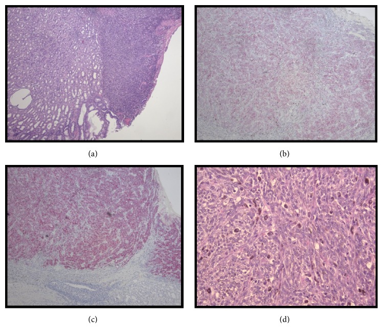
Figure 3.
Histological Specimens of Gastric Melanoma. Histological sections of gastrectomy specimen demonstrating primary gastric melanoma. (a) Right side of the image depicts neoplastic cells that do not invade past the submucosa that still retains normal gastric architecture (H&E x100). (b) Primary gastric melanoma positive stain for Melanin A (H&E x100). (c) Primary gastric melanoma positive stain for HMB-45 (H&E x100). (d) Malignant melanocytic neoplasm producing brown melanin pigment with a cherry red nucleoli and mitotic figures (H&E x 400).

