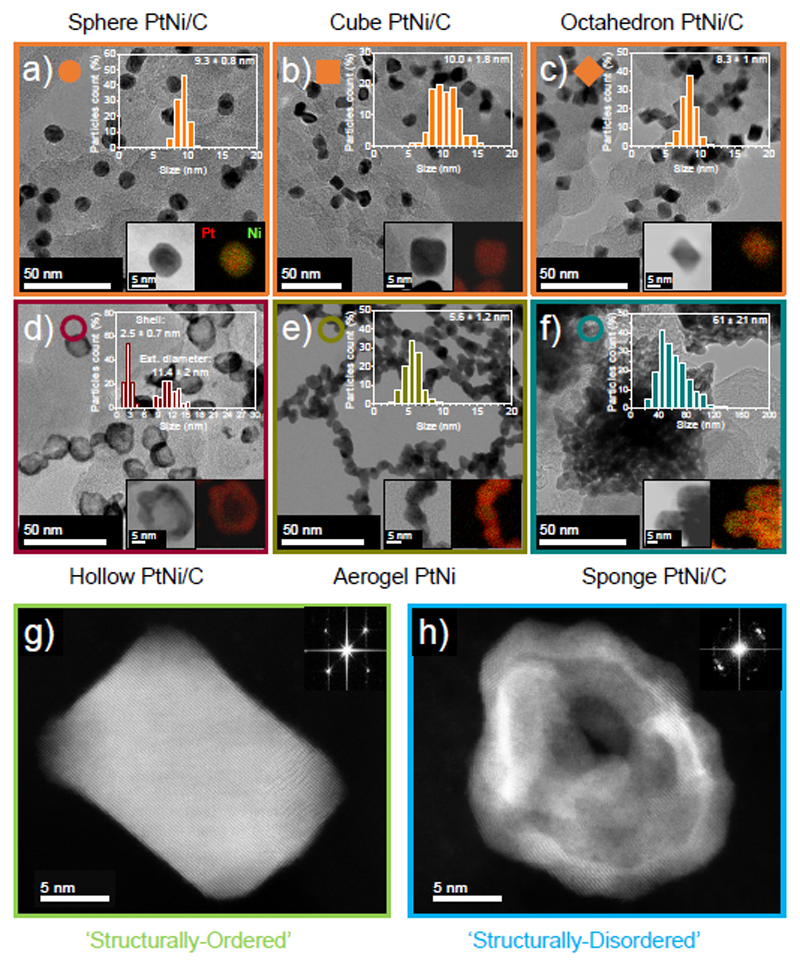
Figure 1. Morphological, structural and chemical characterizations of the various PtNi nanocatalysts synthesized in this study.
Transmission electron microscopy (TEM) image, associated particle size distribution (upper right insert) and scanning electron transmission microscopy coupled with X-ray energy dispersive spectroscopy (STEM/X-EDS) elemental map (lower right insert) of a) Sphere PtNi/C, b) Cube PtNi/C, c) Octahedron PtNi/C, d) Hollow PtNi/C, e) Aerogel PtNi and f) Sponge PtNi/C. High-angle annular dark-field-high resolution scanning transmission electron microscopy (HAADF-HRSTEM) image with insertion of its associated fast Fourier transform pattern of g) Cube Pt/C and h) Hollow PtNi/C. The HAADF-HRSTEM images highlight the monocrystalline nature of the ‘structurally-ordered’ catalyst family (Sphere PtNi/C, Cube PtNi/C and Octahedron PtNi/C) whereas the ‘structurally-disordered’ family (Hollow PtNi/C, Aerogel PtNi and Sponge PtNi/C) features highly polycrystalline nanoparticles. For the Sponge PtNi/C catalyst, the particle size distribution in f) reflects the size of the overall aggregates.

