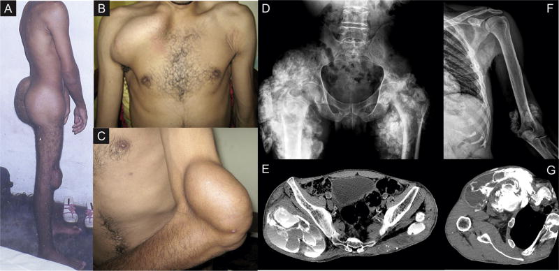
Figure 1.
Metastatic calcifications presenting as firm solid masses over the patient’s (A) gluteal region and knees, (B) right clavicle, and (C) left elbow regions. Image diagnostic methods including radiographs and computed tomography revealed giant calcified masses in the (D) right hip, (E) thigh, (F) left elbow, and (G) near the right clavicle, as well as vascular calcifications.

