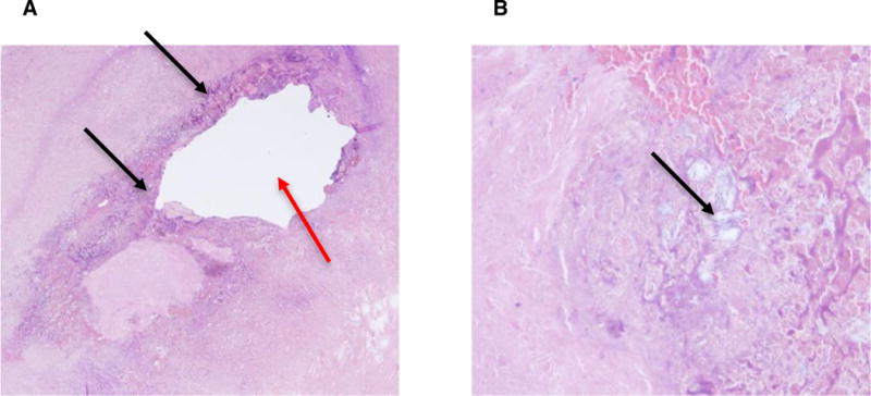
Figure 2.

(A) Hematoxylin and eosin staining shows near circumferential granulomatous inflammation surrounding the previous catheter tip site (red arrow) and multifocal deposition of nonpolarizable precipitated foreign material (black arrows). (B) Higher power view of the nonpolarizable, precipitated foreign material (black arrow).
