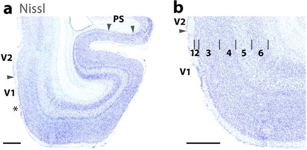
Figure 4.

The laminar characteristics of areas 17 (V1), 18 (V2), and Prostriata (PS) in a horizontal brain section stained for Nissl substance. (a) A lower magnification photo of ventromedial V1 (17) and V2 (18). Arrows mark the 17–18 boundary and the 17/prostriata boundary (PS). A transition zone (marked by asterisk) is apparent in area 17 near the area 18 boundary where some of the laminar features of area 17 are less apparent. Prostriata (arrow heads) is also prominent in the section. (b) A higher magnification of the laminar pattern of area 17. Note the sublaminar pattern in layer 4. Scale bar = 1 mm.
