Figure 1.
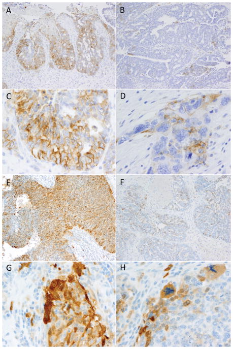
Examples of PD-L1 and IDO tumoral expression patterns in high grade serous ovarian carcinomas. Tumoral PD-L1 expression was seen in a 29% of high-grade serous ovarian carcinomas and never exceeded 10% overall positivity. The case illustrated in plate A had 10% positivity which was concentrated in the focus pictured here; the remainder of the tumor was predominantly PD-L1-negative. In contrast, most positive cases showed a pattern similar to the case illustrated in plate B, with only scattered patches of expression. Crisp membranous staining was required for positivity, as is illustrated in C-D, as was the exclusion of macrophage staining based on comparison with the CD68 stain. IDO expression was more common with 58% of cases showing tumoral staining. Occasional cases showed up to 25% positivity, as pictured in plate E; however, as was observed with PD-L1, such strongly positive swathes of tumor were admixed with larger negative areas. The most common pattern of expression was patchy positivity as pictured in plat e F. Clear cytoplasmic staining as depicted in G–H was required for diagnosis, as was exclusion of macrophages using the CD68 comparison slide.

