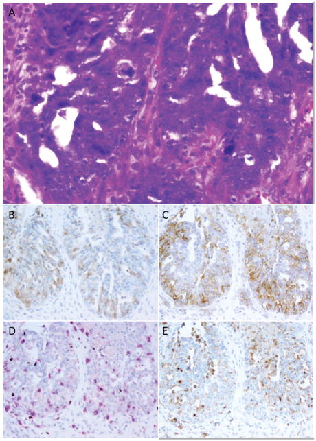
Figure 2. Relationship between PD-L1, IDO, and tumor-associated lymphocytes and macrophages (Example 1).
High-grade serous ovarian carcinomas often showed immune modulatory molecule expression in areas of increased lymphocytic infiltration. This case (A) shows patchy IDO (B) and stronger PD-L1 (C) expression in an area of high density tumor-infiltrating lymphocytes (D; pink=CD8, brown=FOX3p). Some IDO and PD-L1-positive cells correlated with the distribution of CD68-positive tumor-associated macrophages (E) however some tumoral staining was also present for both markers.

