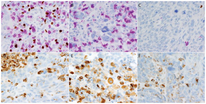
Figure 5.
Examples of tumor-associated lymphocytes and macrophages. Some tumors showed robust infiltrates of both CD8+ (pink) and FOX3p+ (brown) cells (A), while others showed predominantly CD8+ infiltrates (B). Still others showed only sparse lymphocytic inflammation (C). CD68 immunostaining was performed to highlight macrophages in order to facilitate mapping of immune modulatory molecule expression to either tumor cells or macrophages. Plates D–F demonstrate the extent to which CD68-positive macrophages can be subtly admixed among tumor cells, which can complicate assessment of tumoral expression for immune modulatory molecule markers.

