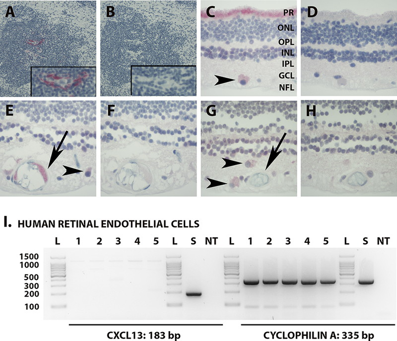
Figure 3.

[A & B] Photomicrograph of immunostained human lymph node showing expression of CXCL13 centrally within the follicle of the tonsil (magnified insert) [A]; serial section immunostained with negative control IgG has no corresponding positive staining [B]. Fast Red with hematoxylin counterstain. Original magnification: 100X. [C - H]. Photomicrographs of human neural retina showing photoreceptor processes [C, ‘PR’] and ganglion cells [C, E & G, arrow heads] staining positively for CXCL13, and vascular endothelium staining positively [E, arrow] or negatively [G, arrow] for CXCL13; matching serial sections immunostained with negative control IgG have no corresponding positive staining [D, F & H]. Fast Red with hematoxylin counterstain. Original magnification: 400X. Abbreviations: PR = photoreceptor layer; ONL = outer nuclear layer; OPL = outer plexiform layer; INL = inner nuclear layer; IPL = inner plexiform layer; GCL = ganglion cell layer; NFL = nerve fiber layer. [I] Agarose gels demonstrating CXCL13 and cyclophilin A (reference gene) RT-PCR products from human retinal endothelial cells isolated from eyes of 5 human cadaver donors, as well as human spleen. Expected product sizes: CXCL13 = 184 bp; cyclophilin A = 335 bp. Lane labels: L = DNA ladder; numbers = human eyes (with each number indicating product from the same individual donor); S = human spleen; NT = no cDNA template.
