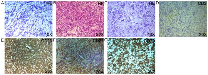
Figure 2.
(A-C) Histopathology of the pulmonary biopsy specimen revealed large numbers of lymphocytes ranging in size from medium to large, with oval or round nuclei containing fine chromatin and scanty cytoplasm (hematoxylin and eosin staining). (D-G) Immunohistochemistry of the pulmonary biopsy specimen revealed positive CD3, CD20, CD21, CD79 expression on the surface of the lymphocytes.

