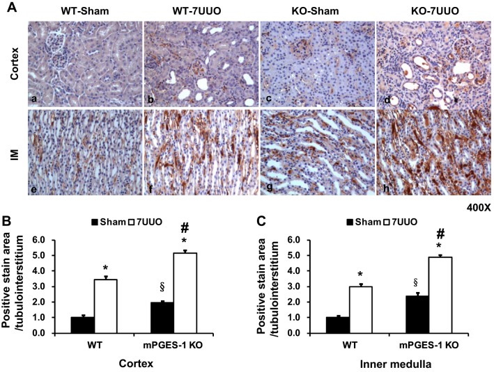
Fig. 8.
mPGES-1 deletion exacerbated UUO-induced increased collagen III staining in kidney cortex and medulla of mPGES-1 WT and KO mice. A: collagen III labeling intensity was significantly increased in obstructed kidneys after UUO. Collagen III labeling in obstructed kidneys of mPGES-1 KO mice with UUO were more extensive and labeling intensity was stronger than WT-7UUO mice. Magnification, ×400. B: quantitative analysis percent of collagen III staining area in mPGES-1 mice on WT-Sham, WT-7UUO, KO-Sham, and KO-7UUO. Statistical analyses were performed by one-way analyses of variance (ANOVA). WT, wild type; KO, knockout. *P < 0.05 compared with Sham groups. §P < 0.05 compared with WT-Sham. #P < 0.05 compared with WT-7UUO.

