Figure 1.
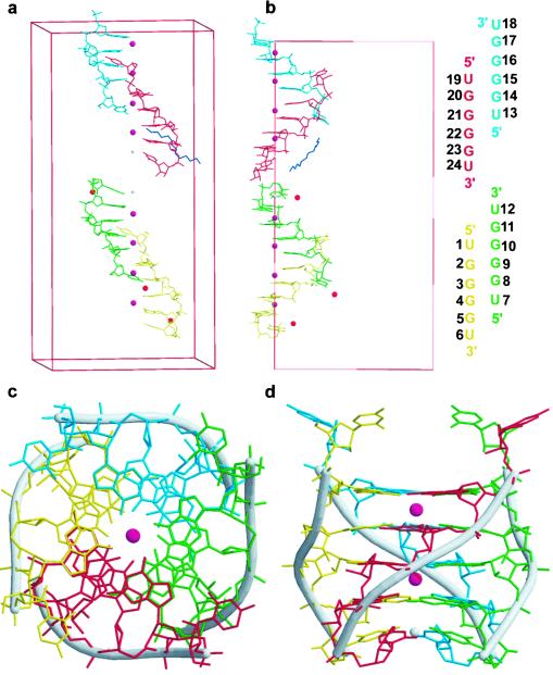
The RNA tetraplex molecule. (a) The four independent RNA strands in the asymmetric unit represented in four different colors: strand 1, yellow; strand 2 green; strand 3 cyan; strand 4 red; Sr2+ ions are in purple and located on the fourfold symmetry axis; Na+ ions are in silver and are also on the fourfold axis; three Ca2+ ions are in general positions and shown in orange; and there is one spermine molecule in general position, which is shown in blue. Sr2+ ions are associated with every other guanine base plane. (b) Another view along crystal b axis. The numbering scheme is shown to the right. (c) Top view of one tetraplex molecule. The Sr2+ ions are sitting on the fourfold symmetry axis, which passes through the helix. The single strand forms a tetraplex with symmetry-related strands. (d) Side view. Notice the 5′-side uridines swing out of the column and the 3′-side uridines are stacked-in and form a U tetrad with a highly tilted angle (≈30°).

