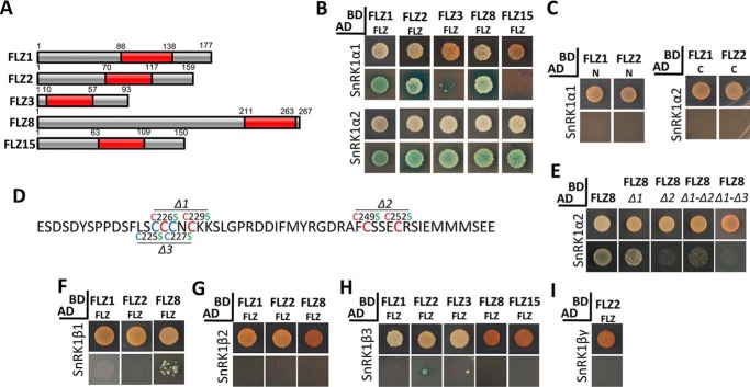
Figure 4.
Role of FLZ domain in mediating the interaction of FLZ proteins with SnRK1 subunits. A, composition of FLZ proteins. The FLZ domain is represented by red box. Number indicates the position of the FLZ domain and other regions in the protein. B, interaction of FLZ domain of FLZ1, FLZ2, FLZ3, FLZ8, and FLZ15 with SnRK1α subunits. C, interaction of N and C termini of FLZ1 with SnRK1α subunits. D, FLZ domain of FLZ8 indicating the position of Δ1, Δ2, and Δ3 mutations. The cysteine residues that form the CX2CX18FCSX2C signature zinc finger motif are indicated by red, and the nonconserved cysteine residues are indicated by blue. E, interaction of FLZ8 and mutated FLZ8 (Δ1, Δ2, Δ1-Δ2, and Δ1-Δ3) with SnRK1α2 subunit. F, interaction of FLZ domain of FLZ1, FLZ2, and FLZ8 with SnRK1β1 subunit. G, interaction of FLZ domain of FLZ1, FLZ2, and FLZ8 with SnRK1β2 subunit. H, interaction of FLZ domain of FLZ1, FLZ2, FLZ3, FLZ8, and FLZ15 with SnRK1β3 subunit. I, interaction of FLZ domain of FLZ2 with SnRK1βγ subunit. The SnRK1 and FLZ constructs were prepared in AD and BD vectors, respectively. The cotransformed colonies of Y2H Gold were screened on DDO (upper row in each group) and QDO plates supplemented with X-α-Gal and AbA (lower row in each group).

