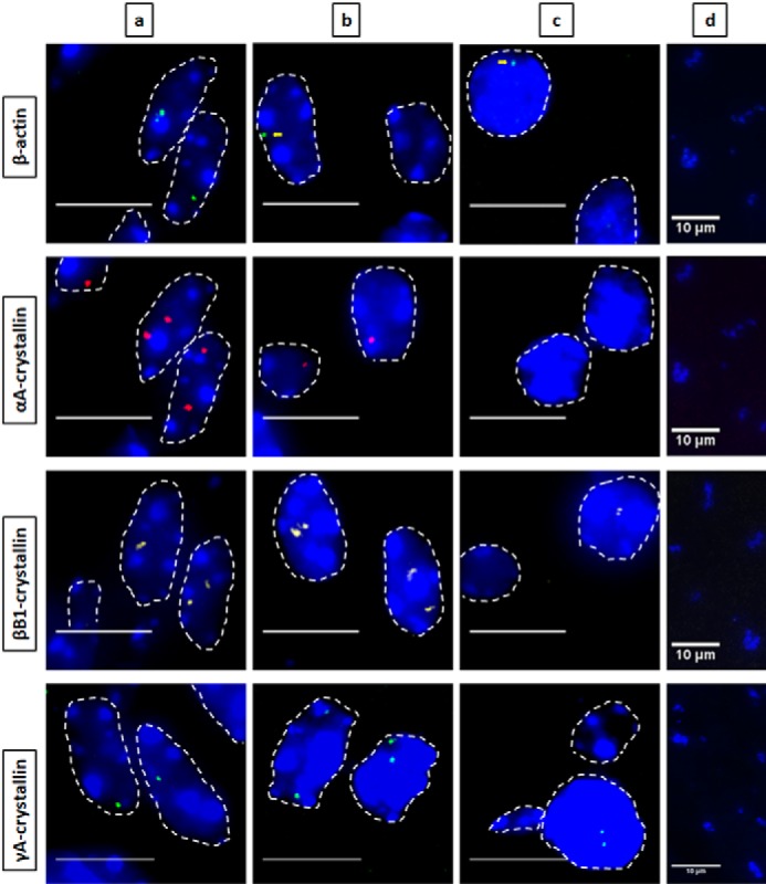
Figure 2.

Nascent transcription sites of various genes during newborn mouse lens fiber cell differentiation show different spatial patterns of transcription for each gene. RNA FISH was performed to show nascent transcription sites of β-actin (Quasar 670, green), αA- (Quasar 570, red), βB1- (Cal Fluor Red 610, yellow), and γA-crystallin (Quasar 670, green) genes at various regions of differentiation in the newborn (P1) mouse lens fiber cells. The areas analyzed are shown in Fig. 1. Regions a, b, and c indicate progressive stages of differentiation throughout the lens tissue with region a being the least differentiated and region c being the most highly differentiated. Region d in this stage of lens development is where denucleation has occurred, and thus, there are no intact nuclei in this region, only degraded pyknotic nuclear remnants. Nuclei were stained by DAPI (blue) and outlined by dotted white lines. A few of the signals are indicated by yellow arrows. Images of nuclei were magnified and adjusted for brightness for viewing this figure. Scale bar, 10 μm.
