
Figure 2.
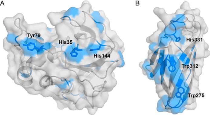
Experimental assessment of the interaction of ScAA10 (A) and ScCBM2 (B) with cellulose nanofibrils. Residues showing normalized amide signal intensities lower than 1 for ScAA10 and lower than 0.1 for ScCBM2 (see Fig. S5) in the presence of cellulose nanofibrils are colored blue. All residues meeting this criterium are visible in the figure, underpinning the clustering of these residues around the putative substrate-binding surfaces (ScAA10, His35, His144, and Tyr79; ScCBM2, Trp275, Trp312, and His331). These experiments were carried out at pH 5.5 and 25 °C.