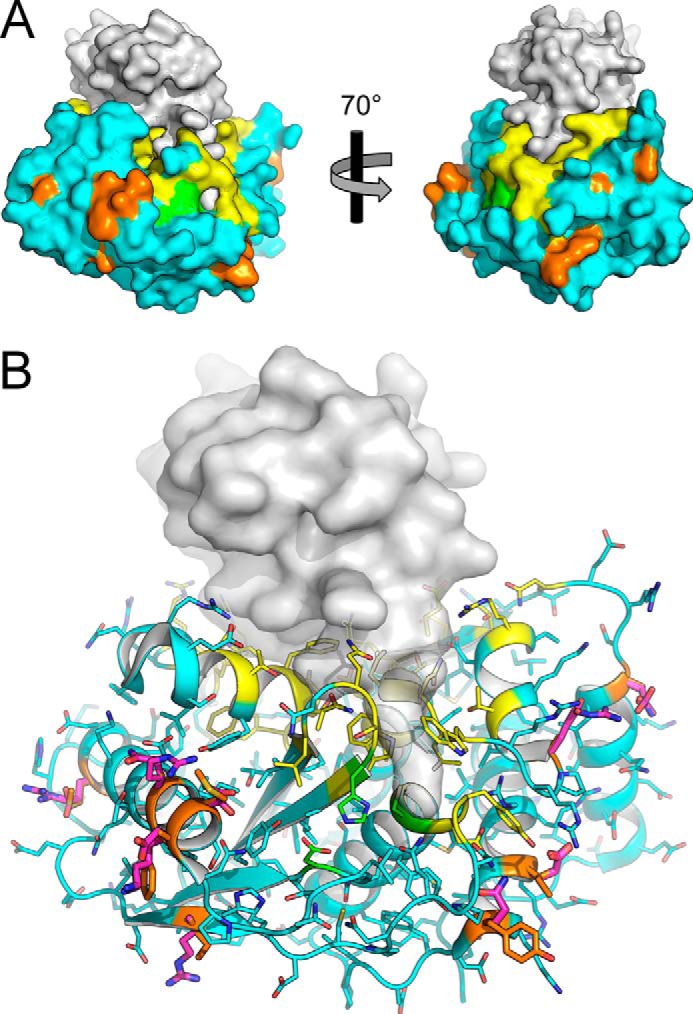
Figure 1.

The SUMO protease Ulp1_WT (colored) is shown in complex with a substrate SUMO protein Smt3 (gray). Nonpolar amino acids of Ulp1_WT selected for computational design are colored orange, and residues that contact SUMO (yellow) or are part of the enzyme catalytic triad (green) were not permitted to change during design; all other residues are colored cyan. Models for Ulp1_WT and Smt3 are from PDB entry 1EUV. A, a molecular surface rendition. B, Smt3 is shown as a semitransparent molecular surface. The Ulp1_WT main chain is shown as a cartoon model, and amino acid side chains are shown as sticks. The side chains of the polar amino acid substitutions from Ulp1_R1–Ulp1_R4 are colored magenta; these and the corresponding side chains of Ulp1_WT are shown in boldface type for emphasis.
