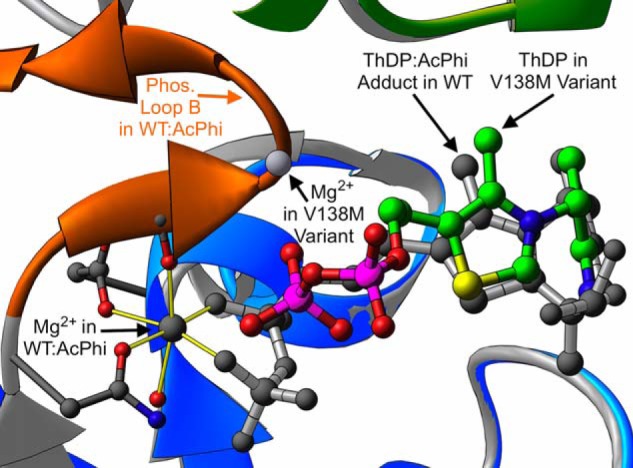
Figure 5.

A depiction of magnesium binding in WT–AcPhi and the αV138M variant. The color coding is the same as described in the legend to Fig. 3. The yellow cylinders indicate the canonical octahedral coordination of the magnesium ion commonly found in ThDP-dependent enzymes. In contrast, in the αV138M variant this binding site is abolished due to the displacement of ThDP's diphosphate tail and the disordering of phosphorylation loop B.
