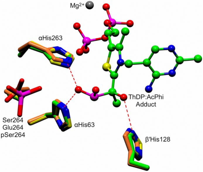
Figure 8.

An active site superposition of the WT–AcPhi E1 structure with the phosphorylation-mimicking αS264E substitution (PDB code 2OZL) and the structure of the WT E1 enzyme phosphorylated at αSer-264 (PDB 3EXH). The ThDP:AcPhi adduct from the WT–AcPhi E1 structure is shown as a ball-and-stick representation. Conserved histidines that interact with and stabilize the substrate analogue are shown as green cylinders (WT-AcPhi), orange cylinders (αS264E substitution), and yellow cylinders (pSer-264). The position of Ser-264, the site of phosphorylation, is indicated.
