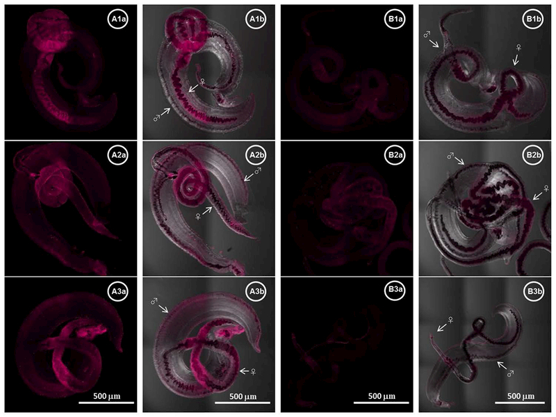
Fig. 4.

Localization of Sm-p80 in S. mansoni adult worms. Representative stitched images of adult worms are shown in panels A (control) and B (vaccinated). The distribution of Sm-p80 in adult worm recovered from three different baboons from control group is shown in A1a–3a respectively while images B1a–3a represent worms from three different vaccinated baboons (magenta). A1b–3b and B1b–3b show a merged of transmitted detector (TD) light and fluorescent Sm-p80. The images were taken using a Nikon T1-E confocal microscopy with a 10× objective and analyzed with NIS software. The stitched images represent a maximum projection intensity derived from a Z-stack. All images acquired with the same laser power and same gain. The arrows indicate the male (♂) and the female (♀) worms respectively.
