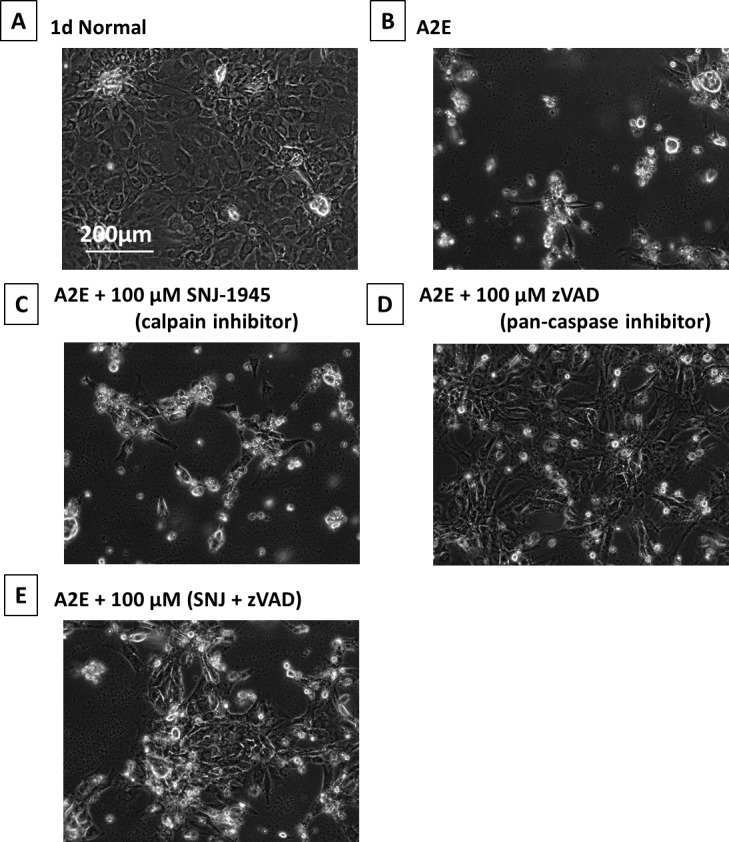
Figure 7.

Phase-contrast micrographs of RPE cells cultured with A2E and inhibitors. (A) 1-day normal, (B) plus 25 μM A2E at 1 day, (C) 25 μM A2E + 100 μM SNJ-1945, (D) 25 μM A2E + 100 μM z-VAD, and (E) 25 μM A2E + 100 μM SNJ-1945 + 100 μM z-VAD. These images were chosen from the most representative experiment in Figure 8 (n = 5).
