Fig. 1.
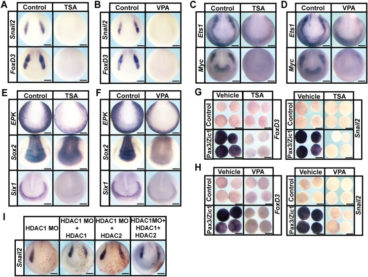
HDAC activity is required for neural crest formation. (A-D) In situ hybridization examining the expression of neural crest factors snail2, foxd3,ets1 and myc following treatment with vehicle or inhibitor [200 nM TSA (A,C) or 20 mM VPA (B,D)]. Embryos were treated at mid-gastrula stages (stage 11) and collected at mid-neurula stages (stage 15). (E,F) In situ hybridization examining expression of epk, sox2 and six1 following treatment with vehicle or inhibitor [200 nM TSA (E) or 20 mM VPA (F)]. (G,H) Explant assay examining snail2 and foxd3 expression in Pax3/Zic1-induced explants treated with vehicle or inhibitor [200 nM TSA (G) or 10 mM VPA (H)]. Explants were cultured alongside sibling embryos grown until late neurula stages (stage 18). (I) In situ hybridization examining the expression of neural crest factor snail2 in embryos after morpholino-mediated knockdown of HDAC1, and rescued with co-injection of HDAC1, HDAC2 or HDAC1+HDAC2 mRNA. Embryos were injected at the eight-cell stage and collected at mid-neurula stages (stage 15). Scale bars: 250 μm.

